Phase Contrast Imaging project
My semester project at ETH with PSI investigated anisotropy in mouse brain phase contrast images to detect neurovasculature.
My semester project at ETH with PSI investigated anisotropy in mouse brain phase contrast images to detect neurovasculature.
Start date: 09.2016
End date: 12.2016
Institutions: ETH Zurich and PSI (Paul Scherrer Institute)
Team: Individual with 1 supervisor
Skills and tools used: image analysis, image reconstruction, phase contrast imaging, micro-computed tomography, OpenCV, Paraview, FIJI, vasculature visualization, image stitching, image correction
Although measuring neurovasculature is of high interest, with developments in different methods,
measuring anisotropy in the brain is limited to date. Developments in phase contrast imaging,
a synchrotron-radiation micro-computed tomography, allow submicron resolution of soft tissue, or
microvasculature. The purpose of this project is to investigate the application of anisotropy analysis
on phase contrast images of a mouse brain sample.
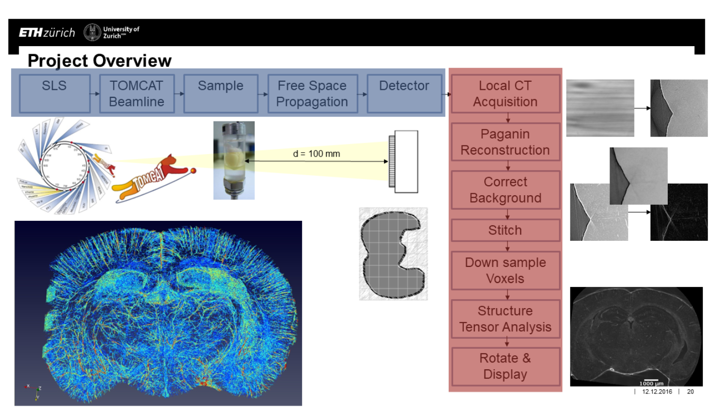
A mouse brain sample was formalin-fixed and perfused with India Ink, as a contrast agent, then imaged
using free space propagation phase contrast imaging at the Paul Scherrer Institute. The sample-to-detector
distance was set to 100 mm and the sample was imaged with a beam energy of 21KeV and a voxel size of 0.65 µm.
Local CT was done with 56 overlapping locations covering almost the complete coronal area of the sample,
taking 2160 projections at each location. The total scan time was ~200 minutes for a volume of ~9 cm. The
captured local CT projections were reconstructed using Paganin’s reconstruction method.
The reconstructed images were cropped, background subtracted and stitched together using FIJI’s Grid/Collection
stitching plugin. To fix the stitching registration and ensure consistent registration between each slice,
a master stitch registration matrix was created and applied to each slice. The resulting data set was divided
into voxels, about 25 µm in size, for anisotropy analysis. Each voxel was Gaussian blurred, with sigma equal to 2,
then the x, y, and z directional gradients were calculated and used to form a structure tensor. To get the
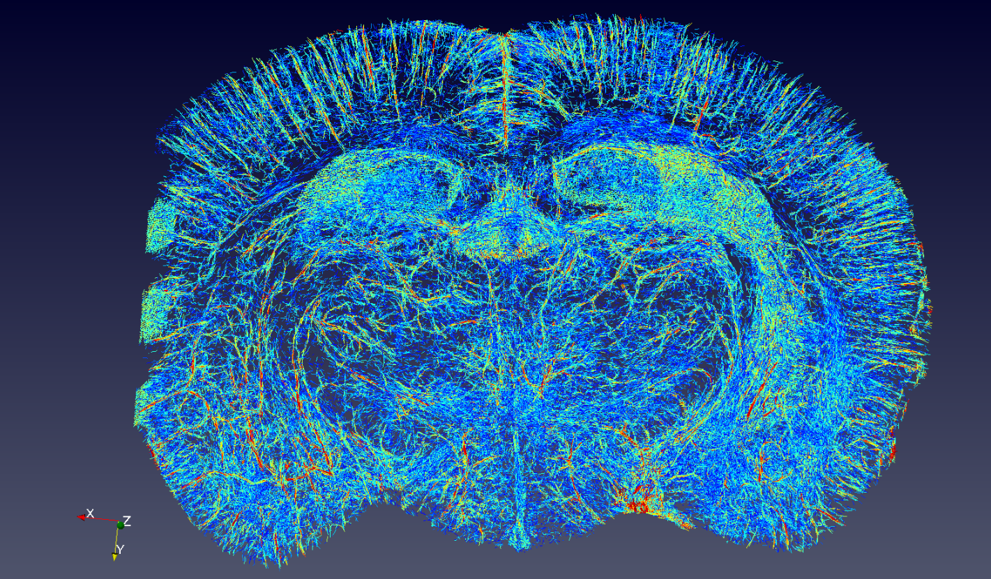
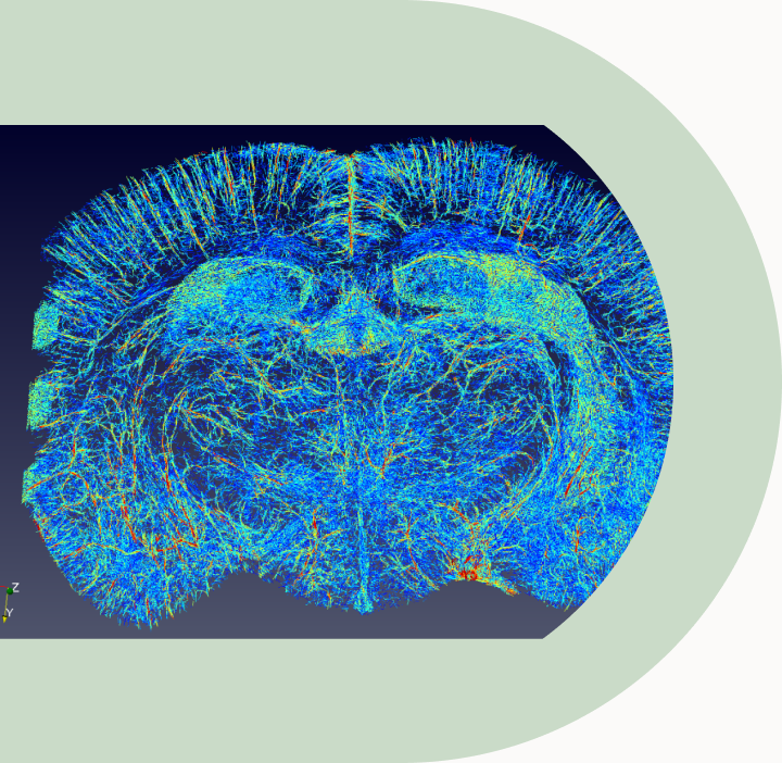
directionality, the eigenvector corresponding to the smallest eigenvalue of the structure tensor was extracted
and saved into a vector map which was masked to remove artefacts and filtered by coherency of 0.5 or greater to
extract anisotropic voxels. The resulting vector map was visualized in Paraview.
The directionality of vessels can be visualized, however limitations exist in regards to the imaging method and
the analysis method. The imaging method is ex vivo with the assistance of a contrast agent and reconstruction
artefacts are present. The structure tensor analysis cannot handle the crossing fibres problem and extracts all
structure features, therefore cannot discriminate only vessels. The next steps of this project include making
improvements to the analysis to enhance vessel extraction, applying tractography in efforts to isolate vessel
connectivity, and correlating the data to other modalities, pathologies and techniques to observe what additional
information or confirmations can be gathered.
In conclusion, submicron resolution phase contrast images were acquired and analysed with anisotropic structure
tensor analysis. Further work is required to refine the analysis but the application of anisotropy to extract
neurovasculature can be a promising technique for visualization.